Non-invasive imaging of atherosclerosis review in EHJ

A very comprehensive review from an eminent group of authors appeared in EHJ recently.

Non-invasive anatomic and functional imaging of vascular inflammation and unstable plaque

It covers:

  1. Pathobiology of plaque, including lipid accumulation, oxidation, inflammation, matrix breakdown, apoptosis and calcification (both macro- and micro-).

  2. Imaging techniques PET, SPECT, CT, MRI and ultrasound

There were some interesting comments on the increasing use of FDG PET imaging for detection of inflammation in atherosclerosis : -

Despite these attractions, some issues require resolution before embracing FDG uptake in this regard. Firstly, only limited prospective data correlate FDG uptake, or changes in FDG uptake, with cardiovascular events or altered rates of such complications,42 and we eagerly await the results of larger prospective cohort studies, such as the High Risk Plaque Initiative and BioImage studies."

"In this regard, nuclear agents that image hypoxia, already in use in oncology, may be
useful in imaging atherosclerosis, as hypoxic conditions may prevail in the core of lesions."


I quite agree on point 1 and keep watching this space for point 2!

Asymptomatic Individuals With a Positive Family History for Premature Coronary Artery Disease and Elevated Coronary Calcium Scores Benefit From Statin Treatment: A Post Hoc Analysis From the St. Francis Heart Study

The goal of this study was to evaluate whether individuals with a positive family history for premature coronary artery disease (CAD) and coronary calcium scoring (CCS) above the 80th percentile might benefit from preventive treatment.

Background

First-degree relatives of patients with premature CAD have an increased risk for cardiovascular disease (CVD), whereas events are poorly predicted in these individuals. Surrogate markers, such as CCS, might refine risk scoring. Nevertheless, the outcome of the St. Francis Heart trial, which investigated the effect of atorvastatin 20 mg/day in asymptomatic individuals with CCS above the 80th percentile, did not reach statistical significance.

Methods

We performed a post hoc analysis on the database of the St. Francis trial to assess efficacy of treatment with atorvastatin 20 mg/day in those with CCS above the 80th percentile and presence (n = 543) or absence (n = 462) of a positive family history for premature CAD. All participants received aspirin 81 mg/day. Primary outcome included coronary death, myocardial infarction, coronary revascularization, stroke, and arterial surgery.

Results

A total of 1,005 individuals, with a mean age of 59.0 ± 5.9 years and a median absolute CCS of 370 Agatston units (interquartile range: 183 to 662) participated in the trial. After a follow-up of 4.3 (interquartile range: 3.5 to 4.5) years, 7.2% of the treated individuals with a positive family history had a cardiovascular event versus 12.5% of the placebo group (hazard ratio [HR]: 0.55; 95% confidence intervals [CI]: 0.31 to 0.97; p = 0.040). This is comparable with a number needed to treat of 18.9. In individuals without a family history, events were minimally reduced: 6.6% in the treated versus 6.8% in the placebo group (HR: 1.04; 95% CI: 0.51 to 2.13; p = 0.912).

Conclusions

The combination of a positive family history and CCS above the 80th percentile identifies a subgroup within the primary prevention population that receives greater benefit from statin treatment than the population at large. These results have important implications for future guidelines concerning individuals with a positive family history for premature CAD.

Via Journal of the American College of Cardiology: Cardiovascular Imaging

Thoughts? Blunt tool?


Chocolate and CV risk - small and dark is best

For those that celebrated Easter with chocolate Eggs, a timely study in the European Heart Journal.


This large, observational study of over 19,000 Germans had a mean follow-up of 8 years. The authors (Buijsse and colleagues) demonstrated that those eating the most chocolate had fewer MI and strokes than more modest consumers (by 39%). Participants were free from cardiovascular disease at baseline.


On average, blood pressure measures were 1 mmHg/0.9 mmHg lower in the highest quartile of chocolate consumption versus the lowest.


It must be said, however, that we are talking about small amounts of chocolate here. Those in the highest quartile consumed 7.5g daily compared with 1.7g daily in the lowest quartile.


This work adds to already published data demonstrating that chocolate can lower blood pressure and improve both endothelial and platelet function.


It's thought that the active ingredient is the cocoa - a flavonoid - also present in red wine, citrus fruits and tea.


Of course the downside is that chocolate is largely sugar - which equals calories - and so must be consumed in small amounts to get the CV benefit.


And it's the expensive stuff, with the highest cocoa content, that would be expected to deliver the most protection.


Sadly, this blog post is not endorsed by Cadbury's, Mars or Godiva.


A larger, more recent meta-analysis is described here.


Any thoughts on this issue? Should we file this under the "All things in moderation" category?


Mcdonald large kid 750701


Imaging atherosclerosis presentation - Barcelona November 2011

Here's a link to the PDF of a talk I gave today in Barcelona at a vulnerable plaque conference, hosted by Valentin Fuster.

What do you think - is it even worthwhile looking for high-risk plaques given that IVUS-VH shows us so many of them?

http://www.evernote.com/shard/s3/sh/cbbc7b18-7871-478b-85a0-f3f934de3cd3/832d02e0abcfb85cdd8d647b95253b2c

 "Detection of the High Risk Atherosclerotic Plaque - The role of PET/CT Imaging".


Aberrant right coronary artery - what next?

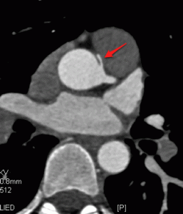
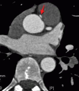
Our hypothetical patient is a 30 year old male construction worker who complained of chest pains at rest, had inferior ST depression and elevated troponin. He settled with medical therapy.

X-ray angiography revealed smooth coronaries without atherosclerosis, but an aberrant RCA.

Subsequent cardiac CT revealed RCA arising from the left coronary sinus of the aorta, but with a separate origin to the left main artery (Image 1). The RCA course was between the ascending aorta and the pulmonary artery. initially the calibre was small (Image 2), widening out once it was in the 'normal' position (Image 3).

The working diagnosis was inferior NSTEMI secondary to compression of the RCA.

What are your views on therapy for this patient?

Is surgery needed? If so, which operation ? Re-implantation of the RCA, or bypass of the RCA?

Is there a role for stenting of the segment lying between PA and Aorta?

Let us know your opinions on how to manage these rare but important patients.


ESC Wrap Up - The End Of The Road For Warfarin In AF?

I thought I'd finish the coverage of ESC by discussing briefly 2 studies that were part of the congress and asking for your opinion.

First, there were further data released from the RE-LY study, covered in depth over at HeartWire - here. This study compared a new oral anticoagulant, dibigatran, head-to-head with warfarin in patients with atrial fibrillation. The results showed that the new drug was non-inferior at low dose and superior at high dose in reducing the number of strokes in the population studied, regardless of the INR control achieved with warfarin. This was a post-hoc analysis from the main study of over 18,000 subjects.

Second, today, there was new data from a competitor product, apixaban, in the same patient group although not directly against warfarin, but against aspirin. The new agent was successful at lowering risk of stroke and systemic embolus (by 34%) without excessive bleeding risk. Head to head studies against warfarin are underway (ARISTOTLE study - reports in 2011).

Is the writing on the wall for warfarin in atrial fibrillation? We don't yet know how the newer drugs will stack up against warfarin for those with rheumatic heart disease and AF. But the ease of use and low bleeding rates will certainly be very attractive to clinicians once the drugs come to the marketplace. Presumably more patients will receive these drugs than currently get warfarin, because of the perceived problems (bleeding, monitoring, drug interactions) with warfarin use.

What do you think? Are you eagerly awaiting warfarin replacements? Or is more evidence required?


Day 4 - ESC

As a change, I thought I'd cover this Day in Stockholm using Twitter feeds. This will give you multiple opinions from a cross section of delegates. Simply click on the links to read more!


Safety of 2 intravenous heparin doses with PCI in ACS patients treated with fondaparinux - here.


ISAR REACT 3A: Reduced dose of unfractionated heparin in patients undergoing percutaneous coronary interventions - here.


Today's hotline press releases - here.


Nurses can reduce the risk of recurrent complications in heart patients: results from the RESPONSE trial - here.


REACH: Comparative Determinants of 4-Yr CV Event Rates in Stable Outpatients at Risk of or with Atherothrombosis - here.


A pill costing less than £1.50 a day has the potential to save thousands of heart failure patients - here.


Hot arteries: Marion Hoffman, Chicago - Vascular muscle cell factor implicated in aortic aneurysms - here.


Future of myocardial perfusion imaging - here.


Day 3 ESC

A very well attended session at the ESC today concerned novel approaches to imaging the vulnerable plaque.

We heard from Zahi Fayad (New York), Jagat Narula (Orange, Ca) and Beat Kaufmann from Basel in Switzerland. I gave an over-view of vascular PET imaging, both with FDG and novel targeted ligands against plaque macrophages.

The session tried to communicate not only the state of play in 2010, but also to predict what may be around the corner for imaging. There seems to be a move towards more platform integration, with the appearance of combined PET/MR hardware. This will allow high sensitivity/high resolution imaging of the artery wall. I guess this will have large impact in brain imaging and likely also in cardiology for myocardial imaging.

For CT, as well as a move to lower radiation dose, Zahi Fayad talked about multi-color CT, something that holds great promise in separating out the various plaque elements, to a much greater extent than currently feasible with single energy imaging with iodine contrast agents. This work was recently published in Radiology (Cormode D et al - see image below).


We also heard a great review of ultrasound imaging of vulnerable plaque, using targeted microbubbles against selectins and adhesion molecules. Whilst very exciting, the field is confined right now to small animal models of disease, and to epitopes expressed on endothelial cells. But there is promise of eventual human translation.

What caught your eye in Stockholm today?