
An interesting concept - a journal that brings research to life using film. Take a look at the first few minutes of a film describing how to do PET/CT scans for vascular inflammation.
Vulnerable plaque imaging comes a step closer
We published a paper in the Lancet last week.
It is the result of a scientific collaboration between Edinburgh and Cambridge Universities. Funding was provided by the British Heart Foundation and the Chief Scientist's Office in Scotland.
The project concerns using PET imaging with NaF to detect atherosclerotic plaques that are in a state of vulnerability to rupture.
Firstly, NaF successfully identified most of the plaques that had caused recent MI because they were brighter after PET imaging than bystander lesions.
We then used IVUS to clarify, in a stable angina cohort, the phenotype of NaF-avid plaques. We also confirmed using immunohistochemistry that NaF binding occurred in areas of both high calcium turnover and inflammation.
The reason that the study is exciting is that it sets the scene for this imaging to be used prospectively, perhaps in high-risk individuals with lesions in several arteries, where it might direct therapy to the most "at-risk" plaques. This might be intensive statin therapy, or even a direct plaque intervention of some type.
We still don't know whether high-risk plaque detection and treatment will alter the natural history of the disease or how cost effective such an approach might be. But it has a major advantage of being non-invasive and relatively low in radiation - about half the dose of a nuclear perfusion scan. There is also no fasting needed before scanning, and NaF is widely available.
The publication received a great deal of media coverage:
British Cardiovascular Society Annual Meeting Preview
Imaging is a theme that runs throughout the conference. Although concentrated within the Imaging Track, there are also sessions and presentations on imaging topics within both the CS/TR and E4R Tracks.
This year, within the Imaging Track, we have sessions dedicated to several pathologies that can sometimes be overlooked, either because they are hard to diagnose or challenging to treat. Disease of the right heart fits this description. We have therefore planned a session that will show how imaging can help the clinician improve their diagnostic accuracy and risk stratification ability. We will discuss both intrinsic conditions of the right heart, as well as its secondary involvement in left-sided heart disease. A partner session will explore how to diagnose and manage pulmonary hypertension. This will include talks on its pathology and the latest diagnostic classification. Presentations on the management of pulmonary hypertension will cover both medical and surgical approaches to the disease.
Another sometimes under-appreciated condition is ‘functional’ mitral regurgitation. In this session, we will discuss its aetiology, and how multi-modality imaging can help both to diagnose and suggest when intervention is needed. We will hear about contemporary management strategies, including how new device options might fit alongside conventional surgical approaches.
We have an entire session dedicated to the pragmatic use of intravascular coronary artery imaging in the catheter laboratory. Here, we will discuss the clinical roles of intravascular ultrasound and optical coherence tomography. Bridging the research and clinical spaces, we will be joined by renowned Harvard cardiologist and entrepreneur Dr James Muller who will educate us about hybrid catheter-based imaging technologies that provide integrated anatomical and functional plaque data. Dr Muller is an expert in using near-infrared spectroscopy for identifying vulnerable plaque.
Imaging is now widely applied in multi-centre trials. In a session on Wednesday, our speakers will discuss the results of several recent large imaging trials. These will include nuclear and echo imaging within the extended STICH study, the latest multi-centre cardiac CT and MR studies, plus how imaging can help decide the best way of revascularising diabetic patients with coronary artery disease.
The ever-popular ‘Read with the Experts’ session is back once again. Do come along and test your image interpretation skills in real-life clinical scenarios presented by multi-modality imaging experts. CT, echo, MR and nuclear imaging approaches will all be on display in one of the most popular sessions of the conference.
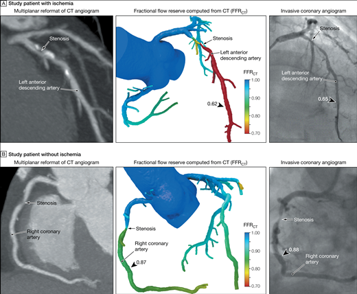
RIPCORD: FFR during diagnostic angio may radically alter treatment plans
Via theheart.org, an intriguing pilot study from Southampton, UK.
Nick Curzen, PI of the trial -
If a randomized trial shows the same results as RIPCORD, that may well be the end of non-interventional cardiologists doing diagnostic angiography
Or how about testing non-invasive FFR using CT angiography - and obviate the need for anyone to do diagnostic angiography?
Advances in Molecular Imaging: Plaque Imaging
Here’s the abstract of a review we published recently in Current Cardiovascular Imaging Reports:-
Recent advances in nuclear plaque imaging aim to achieve noninvasive identification of vulnerable atherosclerotic plaques using positron emission tomography (PET) with 18F-fluorodexoyglucose (FDG) and novel tracers targeting molecular markers of inflammation and other active metabolic processes.
Nuclear imaging of atherosclerosis has been demonstrated in multiple vascular beds, including the carotid, aorta, peripheral and coronary arteries—but significant challenges remain, especially for coronary imaging. The advantage of PET over other molecular imaging modalities is its superior sensitivity, however, low spatial resolution means that images must be co-registered with computed tomography (CT) or magnetic resonance imaging (MRI) for precise anatomical localization of the PET signal.
Such hybrid techniques provide the best hope for early detection of prospective culprit lesions—which may, in the coronary vasculature, appear falsely low-risk using conventional coronary angiography or stress imaging.
Current hot topics in nuclear plaque imaging include the use of FDG-PET for therapeutic monitoring in drug development, identification of imaging biomarkers to evaluate cardiovascular risk, and the development of novel tracers against an array of biologically important markers of atherosclerosis.The purpose of this article is to review these recent advances in nuclear plaque imaging.
The full article is available here, behind a paywall. I can send reprints as PDFs on request.
Learning cardiac CT
There are some great resources out there for learning cardiac CT. One particularly good one is provided by Johns Hopkins radiologist Dr. Elliot Fishman at CT is us.
Here are a couple of his videos to get you started:
British Heart Foundation's Reflections of Research Competition
A video that we submitted to the British Heart Foundation's "Reflections of Research" competition has won in the best video category.
We made this video to highlight the variety of imaging methods that we have at our disposal to image atherosclerosis and its consequences.
Some more coverage on the Cambridge BRC website and the Addenbrooke's Hospital homepage.
Imaging of progression of carotid atherosclerosis - case unproven
A study I recently read in the Lancet reports results of an individual patient data meta-analysis, which show that cIMT progression is not associated with incident myocardial infarction, stroke, or vascular death in the general population (hazard ratio 0·98, 95% CI 0·95–1·01, adjusted for age, sex, mean common carotid artery intima-media thickness, and vascular risk factors).
A single measure of carotid IMT may be useful for predicting risk in asymptomatic intermediate risk adults, although far less so than calcium scoring by CT according to the most recent guidelines.

My PhD thesis now online
I just took advantage of Cambridge's online repository service for research documents. I uploaded my PhD thesis from 2003 - 'Imaging atherosclerosis using FDG PET'. In it, I describe the first prospective study if FDG PET for measuring real-time inflammation in the carotid arteries.
Feel free to download it by visiting this page.
Imaging Early Atherosclerosis
My talk at ESC 2012 in Munich is now available for watching via webcast if you're so inclined.